
Clinical variant interpretation sits at the intersection of genomic science and clinical decision making. For clinicians and bioinformaticians working in precision oncology, the ability to classify variants and translate them into actionable insights is essential. This primer walks through the evolution of variant interpretation—from the early days when terminology was ambiguous to the modern era of consensus guidelines—highlights the key differences between germline and somatic interpretation, outlines best practice approaches and looks ahead to how emerging technologies will reshape the field.
1. A brief history
Early approaches and the pre ACMG/AMP era
Until the mid 2010s, laboratories used inconsistent terminology when describing sequence variants. Terms such as “mutation” and “polymorphism” were used loosely to convey pathogenicity, and qualifiers like “possibly” or “probably” were added to indicate uncertainty. The only widely adopted designation was “variant of uncertain significance” (VUS) (Rehm, 2017). Because there were no standardized rules for weighing evidence, a single paper could be cited to support pathogenicity even when it provided little statistical evidence. Variants deposited in resources like the Human Gene Mutation Database (HGMD) or OMIM were often assumed to be pathogenic without rigorous scrutiny.
As next generation sequencing (NGS) expanded the number of variants identified, laboratories struggled to keep up. It became clear that the community needed shared criteria and a common vocabulary to harmonize variant interpretation.
The ACMG/AMP guidelines and adoption
In 2015, the American College of Medical Genetics and Genomics (ACMG) and the Association for Molecular Pathology (AMP) published detailed standards for interpreting sequence variants. The document defined 28 evidence criteria (e.g., PVS1, PS2/PM6, PM3, PP1, BA1, BS1) across five strength categories (very strong, strong, moderate, supporting and stand alone) and provided rules for combining these criteria to reach one of five classifications: pathogenic, likely pathogenic, VUS, likely benign or benign (Harrison et al., 2019). These guidelines introduced a common vocabulary and have been adopted by >95% of clinical laboratories.
However, even with clear criteria, judgement is required. Initial guidelines intentionally avoided assigning numerical points to each criterion out of concern that users might misinterpret semi quantitative scores as definitive. Nevertheless, subsequent work (e.g., Sherloc and later refinements) introduced point based systems and decision trees to improve reproducibility (Nykamp et al., 2017).
Evolution of somatic variant classification
While the ACMG/AMP standards were designed for germline variants, somatic variants needed a different framework because actionability often depends on tumor type, therapy and evolving evidence. The 2017 joint guidelines from AMP/ASCO/CAP introduced a tiered system for somatic variants (Li et al., 2017):
- Tier I (Level A/B) variants have strong clinical significance—Level A includes biomarkers recognized in FDA approved therapies or professional guidelines; Level B covers biomarkers supported by well powered clinical trials or multiple cohort studies.
- Tier II (Level C/D) variants have potential clinical significance. Level C includes biomarkers with small studies or case reports; Level D covers pre clinical evidence or biologically plausible but unproven associations.
- Tiers III and IV encompass variants of unknown significance and benign alterations respectively.
Because actionability varies by tumor type, the ESMO Scale for Clinical Actionability of molecular Targets (ESCAT) further stratifies variants into six levels (I–V and X). Level I corresponds to targets ready for routine use (e.g., randomised trial showing survival benefit), whereas Level II–V capture investigational or pre clinical evidence, and Level X denotes targets without evidence. ESCAT’s detailed sub levels (I A/I B/I C, II A/II B, etc.) link evidence strength to recommended clinical actions (Mateo et al., 2018).
New consensus recommendations
In 2022, an international team from ClinGen, the Cancer Genomics Consortium (CGC) and the Variant Interpretation for Cancer Consortium (VICC) released oncogenicity guidelines for somatic variants (Horak et al., 2022). These guidelines distinguish oncogenicity (biological driver status) from clinical actionability. Variants are classified as oncogenic, likely oncogenic, VUS, likely benign or benign. Evidence codes (O1–O11 for oncogenicity, B1–B7 for benignity) are weighted numerically. Two or more lines of evidence are usually required to reach a “(likely) oncogenic” or “(likely) benign” call, and evidence is not “stacked” across similar sources (e.g., multiple databases cannot raise the strength of the same code).
The Association for Clinical Genomic Science (ACGS) ratified a UK standard for somatic variant interpretation in 2025. It adopts a points based system inspired by ClinGen/CGC/VICC and the UK Best Practice Guidelines. Canonical driver variants (e.g., BRAF V600E) that are supported by robust functional data automatically receive high scores (O1). Other evidence categories include null variants in tumor suppressor genes (O2/B2) and enrichment in tumor databases (O4). The guidelines emphasize that at least two independent evidence items are needed for classification and recommend resources like the Cancer Gene Census and Cancer Genome Interpreter to determine gene mode of action.
2. Germline versus somatic interpretation
Germline and somatic variants originate from different biological contexts and therefore require different testing approaches and interpretive frameworks (Gray et al., 2018).
- Origin and purpose. Germline variants are inherited and are present in every cell; testing identifies hereditary cancer syndromes and informs family counseling. Somatic variants arise after conception and are confined to tumor tissue; testing seeks biomarkers for targeted therapy or diagnosis.
- Classification frameworks. Germline classification relies on the ACMG/AMP five tier system, often refined for specific genes or diseases. Somatic classification uses tier based systems such as AMP/ASCO/CAP, ESCAT and the ClinGen/CGC/VICC oncogenicity framework.
- Reporting and consent. Germline testing may reveal incidental findings with implications for relatives; patient consent and genetic counseling are essential. When tumor testing identifies variants suggestive of germline origin, laboratories should recommend confirmatory testing and disclose only with appropriate consent. Negative results in therapy relevant genes should also be reported because they impact eligibility for targeted treatments.
- Inheritance vs. tumor biology. Germline variants are inherited at conception, whereas somatic variants may occur at any time during life and often reflect selective pressures within the tumor microenvironment.
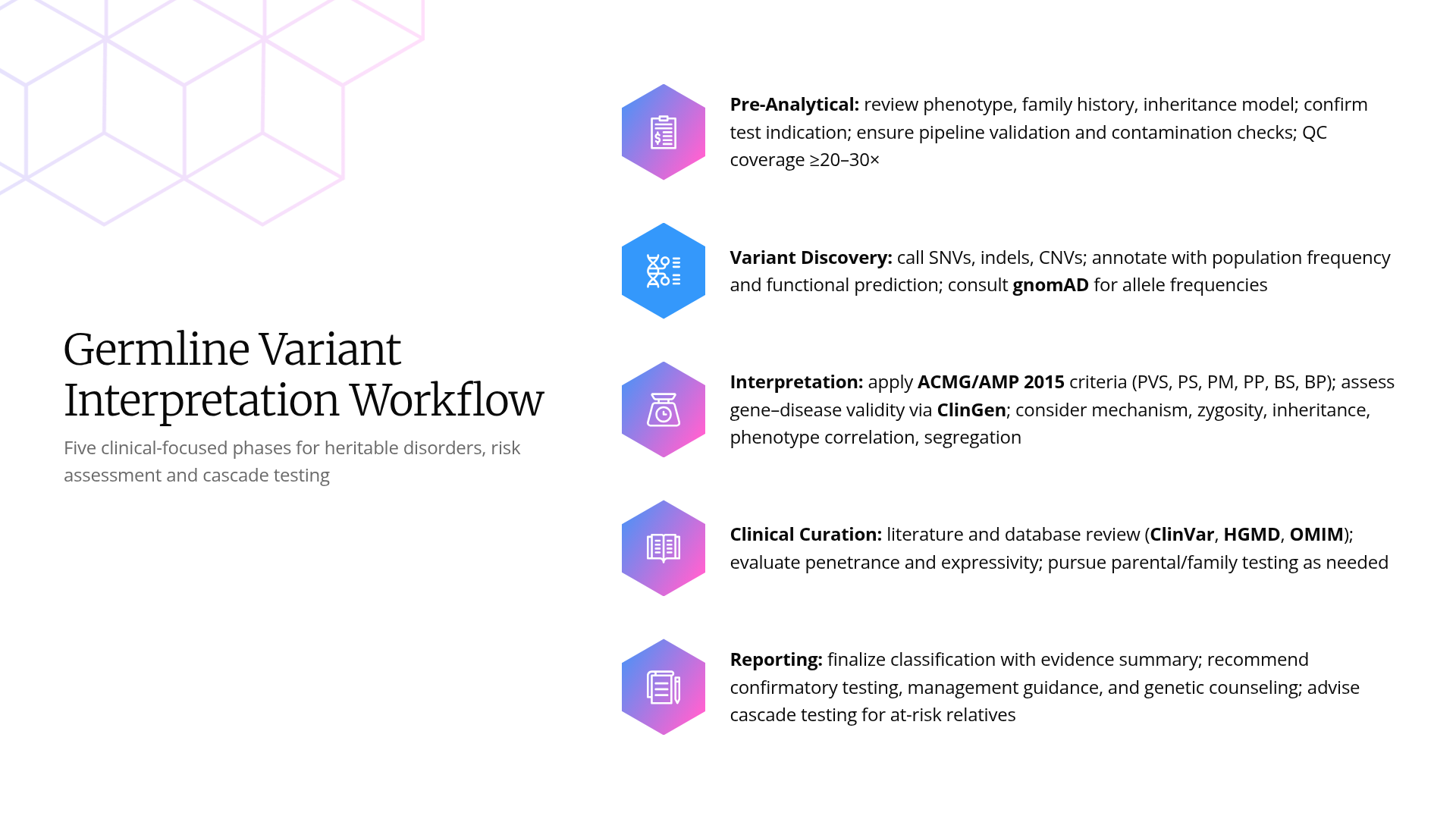
Figure 1. Germline variant interpretation and reporting workflow.
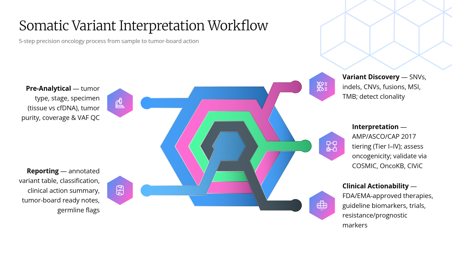
Figure 2. Somatic variant interpretation and reporting workflow.
3. Best practices for variant interpretation and reporting
Bioinformatics pipeline and variant calling
Accurate variant interpretation begins with a robust analytic pipeline. Recent guidelines for clinical NGS recommend:
- Adopting up to date reference genomes (e.g., GRCh38/hg38).
- Using multiple variant callers and cross checking results against truth sets (Genome in a Bottle for germline variants and SEQC2 for somatic variants) to benchmark sensitivity and specificity.
- Containerising analysis environments to ensure reproducibility and facilitate validation.
Variant classification workflow
- Context matters. Begin by reviewing the clinical context: patient’s cancer type, stage, treatment history and family history. Interpretation of the same variant (e.g., EZH2 mutations) can change depending on whether the gene acts as a tumor suppressor or oncogene in the specific tumor type.
- Evidence gathering. Retrieve genomic coordinates in HGVS and GRCh38 notation and consult databases (ClinVar, COSMIC, OncoKB) for known interpretations. For somatic variants, check whether the variant appears on canonical lists (O1) or is enriched in large tumor datasets (O4).
- Apply evidence criteria. Use appropriate frameworks:
- ACMG/AMP for germline variants—apply relevant criteria (PVS1, PM2, PP3, etc.) and combine according to specified rules.
- AMP/ASCO/CAP or ESCAT for clinical actionability—assign tiers based on therapy guidelines, clinical trials and pre clinical evidence.
- ClinGen/CGC/VICC and ACGS frameworks for oncogenicity—use point based evidence codes, require at least two independent lines of evidence and avoid over weighting single sources.
- Interpretation and reporting. Present variants with full gene and protein nomenclature, variant allele frequency (VAF) and zygosity. List clinically relevant variants and VUS, and include negative results for genes relevant to therapy selection (e.g., a KRAS wild type result may qualify a colon cancer patient for anti EGFR therapy).
- Disclose potential germline findings cautiously. When a tumor test reveals a likely germline pathogenic variant (e.g., a BRCA1 truncating mutation), clearly state the possibility and recommend germline confirmation. Only report incidental findings when the patient has consented and the information is clinically actionable.
- Documentation and data sharing. Capture the reasoning behind each classification—including the evidence codes applied and any literature reviewed. Submit interpretations to public databases such as ClinVar to contribute to community knowledge and enable consensus building.
Lessons from real world cases
In our practice, variant interpretation often hinges on nuanced details. Some illustrative scenarios include:
- Subclonal driver mutations. Low frequency BRAF V600E mutations in a melanoma sample may be genuine subclonal events rather than sequencing artefacts; cross checking read directionality and verifying with orthogonal methods can prevent misclassification.
- Unexpected germline discoveries. Tumor sequencing of a colon carcinoma revealed a frameshift mutation in MLH1 with ~50% VAF. The variant triggered ACMG criterion PVS1 and, combined with absence from population databases, was classified as likely pathogenic. Given the VAF and cancer type, we suspected Lynch syndrome and recommended germline testing. The patient consented and subsequent testing confirmed a germline origin, enabling cascade testing for relatives.
- Context dependent oncogenes. A p.Tyr641 EZH2 mutation is oncogenic in follicular lymphoma but may not drive myeloid malignancies. Assigning the correct classification required consulting disease specific guidelines and understanding the gene’s mode of action in each tumor type.
- Actionability vs. oncogenicity. A rare PIK3CA mutation in a breast tumor achieved “likely oncogenic” status under the ACGS framework but fell into ESCAT Level II because evidence for therapeutic benefit came from early phase trials. The patient was eligible for a phase II trial rather than receiving off label therapy.
These cases underscore the importance of combining molecular evidence with clinical context and of communicating uncertainties to clinicians and patients.
4. The future of precision oncology
Emerging technologies promise to make variant interpretation richer and more dynamic. A 2025 review of precision oncology highlights several trends (Brlek et al., 2025):
- Spatial transcriptomics and single cell analysis retain spatial context when mapping gene expression. They reveal interactions between cancer and immune cells, inform immunotherapy design and help identify microenvironment specific vulnerabilities.
- Liquid biopsies—analysis of circulating tumor DNA or other biomarkers—offer minimally invasive monitoring of tumor evolution and therapy response. With improved sensitivity and standardized pipelines, liquid biopsy may enable early detection, minimal residual disease monitoring and timely identification of resistance mutations.
- Multi omics integration combines genomic, transcriptomic, proteomic, metabolomic and epigenomic data. Artificial intelligence tools are increasingly able to identify patterns in these high dimensional datasets, predict treatment response and stratify patients.
- Data driven models and equitable access. The convergence of advanced sequencing technologies, computational frameworks and cloud based data sharing will produce sophisticated models capable of predicting disease trajectories and optimizing individualized regimens. As technological costs fall and regulatory frameworks mature, precision oncology must prioritize equitable access to testing and targeted therapies.
Conclusion
Variant interpretation has evolved from a patchwork of inconsistent terminology into a rigorously codified discipline. The 2015 ACMG/AMP guidelines established a common framework for germline variants, and subsequent developments—including tiered somatic guidelines, ESCAT and the ClinGen/CGC/VICC oncogenicity system—have provided structure for somatic and actionable findings. Emerging guidelines like the ACGS framework emphasize point based scoring and context dependent evaluation, ensuring that variant interpretation remains adaptable to new evidence.
Best practice demands a robust analytical pipeline, careful application of evidence criteria, transparent reporting and collaboration across laboratories. As precision oncology embraces spatial biology, liquid biopsy, multi omics integration and artificial intelligence, the interpretive frameworks built over the past decade will serve as a foundation for even more nuanced and personalized cancer care.
References
- Brlek, P., Škaro, V., Hrvatin, N., Bulić, L., Petrović, A., Projić, P., Smolić, M., Shah, P., & Primorac, D. (2025). Advances in Precision Oncology: From Molecular Profiling to Regulatory-Approved Targeted Therapies. Cancers (Basel), 17(21). https://doi.org/10.3390/cancers17213500
- Gray, S. W., Gagan, J., Cerami, E., Cronin, A. M., Uno, H., Oliver, N., Lowenstein, C., Lederman, R., Revette, A., Suarez, A., Lee, C., Bryan, J., Sholl, L., & Van Allen, E. M. (2018). Interactive or static reports to guide clinical interpretation of cancer genomics. J Am Med Inform Assoc, 25(5), 458–464. https://doi.org/10.1093/jamia/ocx150
- Harrison, S. M., Biesecker, L. G., & Rehm, H. L. (2019). Overview of Specifications to the ACMG/AMP Variant Interpretation Guidelines. Curr Protoc Hum Genet, 103(1), e93. https://doi.org/10.1002/cphg.93
- Horak, P., Griffith, M., Danos, A. M., Pitel, B. A., Madhavan, S., Liu, X., Chow, C., Williams, H., Carmody, L., Barrow-Laing, L., Rieke, D., Kreutzfeldt, S., Stenzinger, A., Tamborero, D., Benary, M., Rajagopal, P. S., Ida, C. M., Lesmana, H., Satgunaseelan, L., … Sonkin, D. (2022). Standards for the classification of pathogenicity of somatic variants in cancer (oncogenicity): Joint recommendations of Clinical Genome Resource (ClinGen), Cancer Genomics Consortium (CGC), and Variant Interpretation for Cancer Consortium (VICC). Genet Med, 24(5), 986–998. https://doi.org/10.1016/j.gim.2022.01.001
- Li, M. M., Datto, M., Duncavage, E. J., Kulkarni, S., Lindeman, N. I., Roy, S., Tsimberidou, A. M., Vnencak-Jones, C. L., Wolff, D. J., Younes, A., & Nikiforova, M. N. (2017). Standards and Guidelines for the Interpretation and Reporting of Sequence Variants in Cancer: A Joint Consensus Recommendation of the Association for Molecular Pathology, American Society of Clinical Oncology, and College of American Pathologists. J Mol Diagn, 19(1), 4–23. https://doi.org/10.1016/j.jmoldx.2016.10.002
- Mateo, J., Chakravarty, D., Dienstmann, R., Jezdic, S., Gonzalez-Perez, A., Lopez-Bigas, N., Ng, C. K. Y., Bedard, P. L., Tortora, G., Douillard, J. Y., Van Allen, E. M., Schultz, N., Swanton, C., André, F., & Pusztai, L. (2018). A framework to rank genomic alterations as targets for cancer precision medicine: the ESMO Scale for Clinical Actionability of molecular Targets (ESCAT). Ann Oncol, 29(9), 1895–1902. https://doi.org/10.1093/annonc/mdy263
- Nykamp, K., Anderson, M., Powers, M., Garcia, J., Herrera, B., Ho, Y. Y., Kobayashi, Y., Patil, N., Thusberg, J., Westbrook, M., & Topper, S. (2017). Sherloc: a comprehensive refinement of the ACMG-AMP variant classification criteria. Genet Med, 19(10), 1105–1117. https://doi.org/10.1038/gim.2017.37
- Rehm, H. L. (2017). A new era in the interpretation of human genomic variation. Genet Med, 19(10), 1092–1095. https://doi.org/10.1038/gim.2017.90